Lameness types
Topics
3 min read
Lameness in New Zealand's dairy herds is primarily caused by five types of hoof lesions: White line disease, Sole problems (including bruises, abscesses, and ulcers), Hoof wall cracks, Foot rot, and Digital dermatitis. This page details details the visual appearance, affected claws, and risk factors of these main lameness types. It explains how they occur, particularly focusing on specific points of the cows' feet, and the unique conditions and triggers for each type of lameness. By understanding these, farmers can identify and manage these issues more effectively to ensure the health of their herd.
There are five main types of hoof lesions in New Zealand herds.

The main types of lameness in New Zealand are:
Blue area: White line disease (Above coronary band - White Line disease break out point.)
Green area: Sole
Haemorrhage / bruise (1 or 2)
Absess (2)
Ulcer (1)
➖ Hoof wall crack
⬤ Foot rot
▲ Digital dermatitis
Some lameness is above the claw or the type is unclear/unknown.
See visual examples, what claws are affected and the risk factors for the main lameness types.
What it looks like:

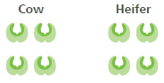
Which foot
Mostly seen in:

How does it happen?
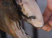
The white line is a weak point in the hoof and if damaged can lead to the white line separating.
At calving the white line weakens and is more susceptible to damage.
Twisting and turning of feet on tracks and yards puts increased pressure on the white line.
Stones can be pushed up into a separated white line resulting in further damage.
If this continues, stone and bacteria will reach the sensitive tissues of the hoof causing pain and infection.
If not treated early and effectively permanent damage to the bone within the hoof can occur, which will increase the risk of the cow getting lame again in the future
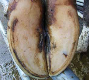
What does it look like?
Reddish/dark brown areas on the sole
Patches can be localised or they can cover large portions of the sole
Often the cow is lame in more than one foot and they are stiff when getting up and walking.
Bruising remains for weeks after the initial damage and may be present along with other sole lesions and WLD, so always check for these other lesions.

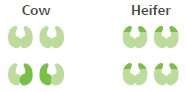
Which foot?

How does it happen?
At calving the tissue below the sole becomes more susceptible to damage (haemorrhage)
From very thin soles.
What does it look like?
Which foot?
How does it happen?
What does it look like?

Which foot?

How does it happen?
At calving the sensitive tissue below the sole is more susceptible to damage which can result in defects in the sole horn
Can be from long periods of standing on concrete e.g. feed pads
What does it look like?
Which foot?
How does it happen?
Damage to the soft tissue between the claws that then grows down as a crack. Risk factors are the same as for foot rot.
Poor conformation of feet e.g. corkscrew.
What does it look like?

Which foot?

How does it happen?
Usually the skin between the claws is broken by a stone, especially under moist hoof conditions.
Bacteria then invade the soft tissue causing an infection.
The onset of foot rot is rapid and will continue for at least a week or until complications set in.
It is a very painful condition.
Digital dermatitis is a highly infectious bacterial skin disease of the feet which is a significant cause of lameness in cattle overseas. It thrives on dirty feet and spreads in dirty conditions.
The disease was discovered in Italy in 1974 and was first identified in five New Zealand herds in 2011. Two years later a further 40 infected herds were discovered. A 2014-15 pilot study conducted in Taranaki found more than 50% of dairy herds tested had some infected animals.
What does it look like?

Which foot?

How does it happen?
Now’s the perfect time to check in, plan, and set up for a strong season. We’ve pulled together smart tips and tools to help you stay ahead all winter long.
Whether you prefer to read, listen, or download handy guides, we’ve got you covered with trusted tools to support your journey every step of the way.


