Types of lameness
Topics
3 min read
Lameness in New Zealand's dairy herds is primarily caused by five types of hoof lesions: White line disease, Sole problems (including bruises, abscesses, and ulcers), Hoof wall cracks, Foot rot, and Digital dermatitis. This page shows what each looks like, which claws are affected, and the main risk factors. Understanding these and how they occur helps farmers identify and manage them more effectively, ensuring the health of their herd.
There are five main types of hoof lesions in New Zealand herds which are:
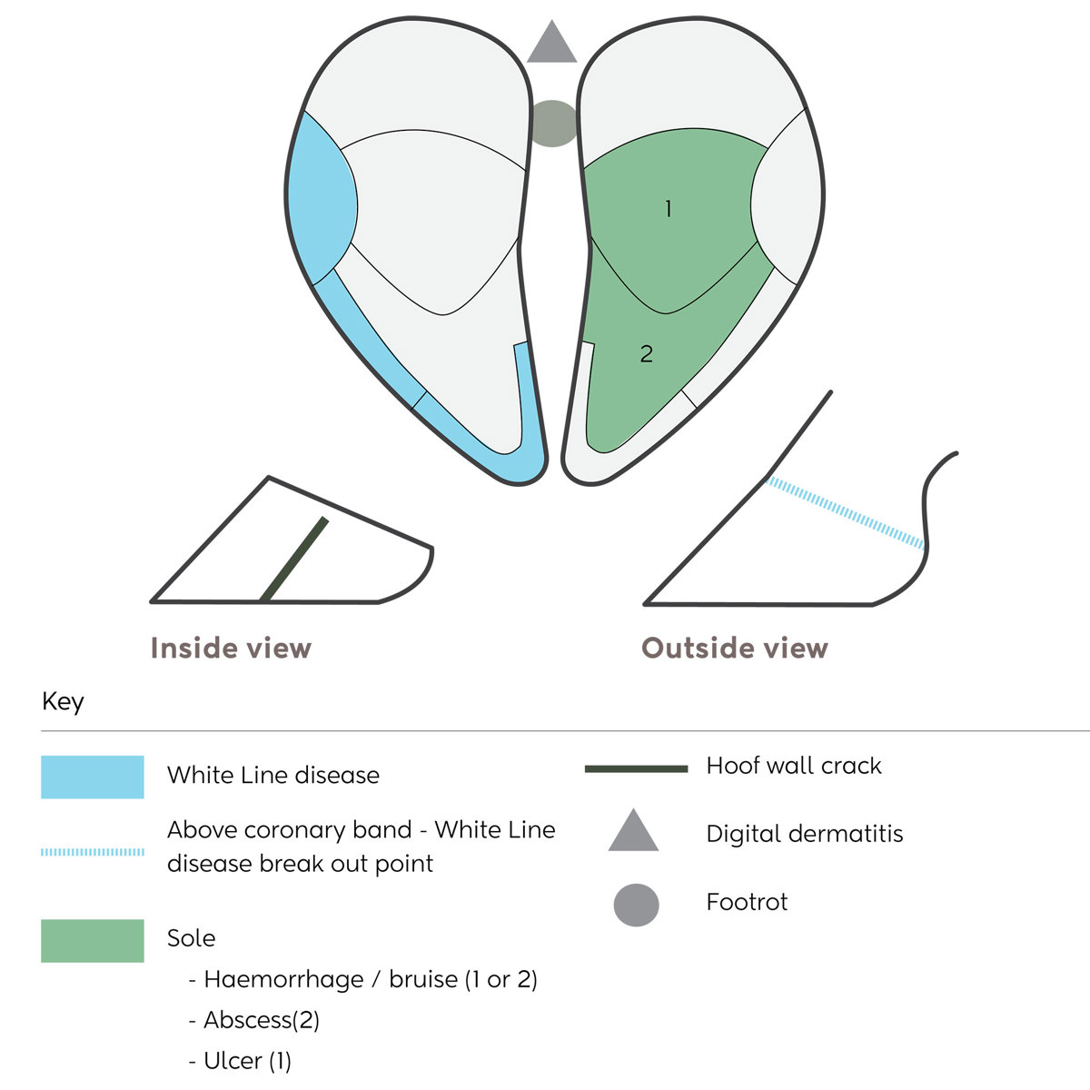


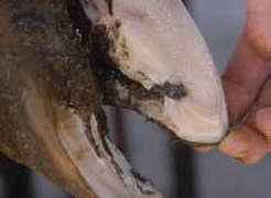
What does it look like?
Where in the hoof?
How does it happen?

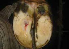
What does it look like?
Where in the hoof?
How does it happen?
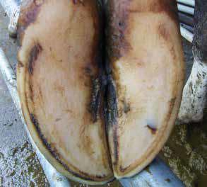
What does it look like?
Where in the hoof?
How does it happen?
What does it look like?
Which foot?
How does it happen?
What does it look like?
Where in the hoof?
How does it happen?
Damage to the soft tissue between the claws that then grows down as a crack. Risk factors are the same as for foot rot.
Poor conformation of feet e.g. corkscrew.
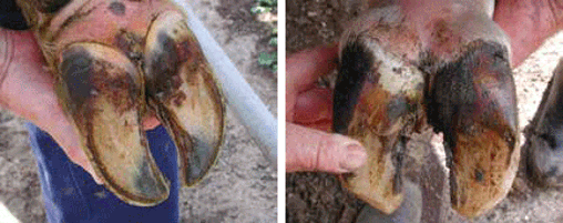
What does it look like?
Where in the hoof?
How does it happen?
Usually, the skin between the claws is broken by a stone, especially under moist hoof conditions.
Bacteria then invade the soft tissue, causing an infection.
The onset of foot rot is rapid and will continue for at least a week or until complications set in.
It is a very painful condition.
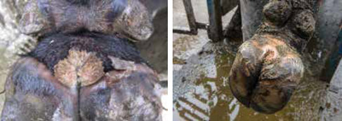
Digital dermatitis is a highly infectious bacterial skin disease of the feet which is a significant cause of lameness in cattle overseas. It thrives on dirty feet and spreads in dirty conditions.
The disease was first identified in five New Zealand herds in 2011 and is becoming more widespread.
What does it look like?
Which foot?
How does it happen?
Now’s the perfect time to check in, plan, and set up for a strong season. We’ve pulled together smart tips and tools to help you stay ahead all winter long.
Whether you prefer to read, listen, or download handy guides, we’ve got you covered with trusted tools to support your journey every step of the way.
Put our proven strategies and seasonal tools to work. Boost production, support animal health and watch your profits hum.
Tools that are backed by science, shaped by farmers and made for this season.
That’s Summer Smarts.
Autumn Smarts brings together the research-backed tools that build resilience and boost performance.


